Field note
U-Net: the architecture built for too little data
Why the encoder-decoder-with-skip-connections shape became the default starting point for biomedical segmentation, and how it was designed specifically around having very few annotated images.
Most computer vision benchmarks in 2015 had thousands to millions of labelled images. Biomedical microscopy almost never does — annotating cell boundaries pixel-by-pixel is slow, and a typical experiment might yield a few dozen usable training images. U-Net was designed around that constraint from the ground up, and the shape it introduced — an encoder that compresses the image down, a decoder that expands it back out, connected by shortcuts across matching resolutions — is still the base architecture that most microscopy segmentation tools, including several referenced elsewhere on this timeline, build on.
The shape explains the name
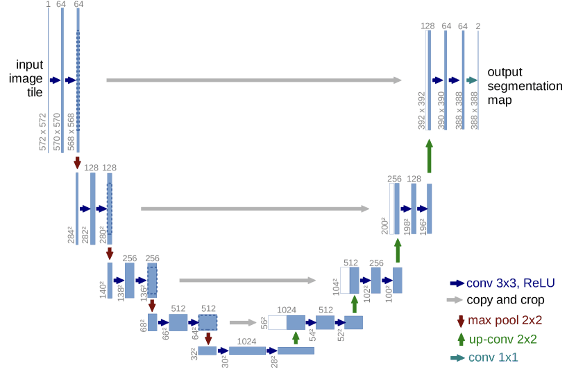
Drawn out, the network looks like the letter U: a contracting path repeatedly downsamples the image, building up an understanding of what is in it at the cost of losing precise spatial detail; an expanding path then upsamples back to the original resolution to decide where each pixel belongs. Left on its own, that round trip would lose the fine boundary detail needed for precise segmentation — so U-Net adds skip connections that copy feature maps directly from each level of the contracting path across to the matching level of the expanding path. The decoder gets both the coarse “what” from deep in the network and the precise “where” preserved from early on, combined at every resolution.
Built for too little data, on purpose
Two design choices target the small-dataset problem directly, not as an afterthought:
- Aggressive elastic deformation as data augmentation — smoothly warping training images to simulate realistic biological variation (tissue isn’t rigid) — let the network learn robust invariances from a training set of only a few dozen annotated images.
- A weighted loss that up-weights the thin background separating touching objects. Densely packed cells that touch are the single most common way naive segmentation fails: without this weighting, the network tends to merge adjacent cells into one blob rather than learning where one ends and the next begins.
Why it became the default
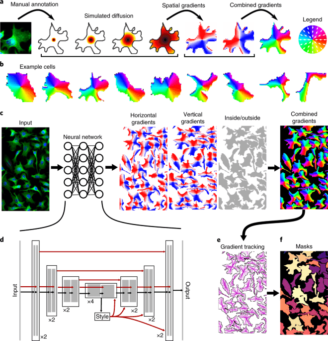
U-Net won the ISBI 2015 cell tracking challenge by a wide margin using a training set that would have been considered far too small for most vision architectures at the time. What generalised beyond that one competition was the shape — encoder, decoder, skip connections — not the specific cell-membrane task it was built for. Cellpose, StarDist, and most other microscopy segmentation tools referenced on this site’s Evolution timeline use some variant of this same encoder-decoder-with-skip-connections backbone, adapted with different output representations (flow fields, star-convex polygons) rather than a different core shape.
Takeaways
- The core idea is a trade: downsampling builds semantic understanding but destroys spatial precision; skip connections give the decoder both, instead of forcing a choice between them.
- U-Net’s augmentation and loss design were built specifically to work with very few labelled images — a constraint that still describes most microscopy experiments.
- What spread through the field was the architectural shape, not the original neuronal-membrane segmentation task — it’s now a general-purpose backbone, not a specialist tool.
- If cells or objects in your images touch or overlap densely, the loss function matters as much as the architecture: a plain per-pixel loss without boundary weighting tends to merge touching instances.
Source
Ronneberger, Fischer & Brox, U-Net: Convolutional Networks for Biomedical Image Segmentation, MICCAI 2015 — paper. See this entry in context on the Evolution timeline.
Continue through the concepts
Related notes
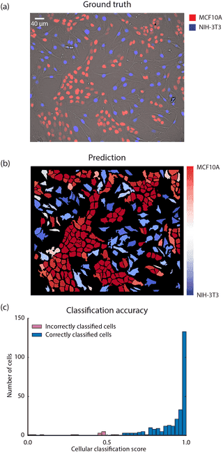 Van Valen et al.: CNNs learn live-cell tracking from ~100 annotated cells
One of the earliest demonstrations that a small amount of manual annotation was enough to train a CNN that out-tracked classical live-cell segmentation methods — the work that grew into DeepCell.
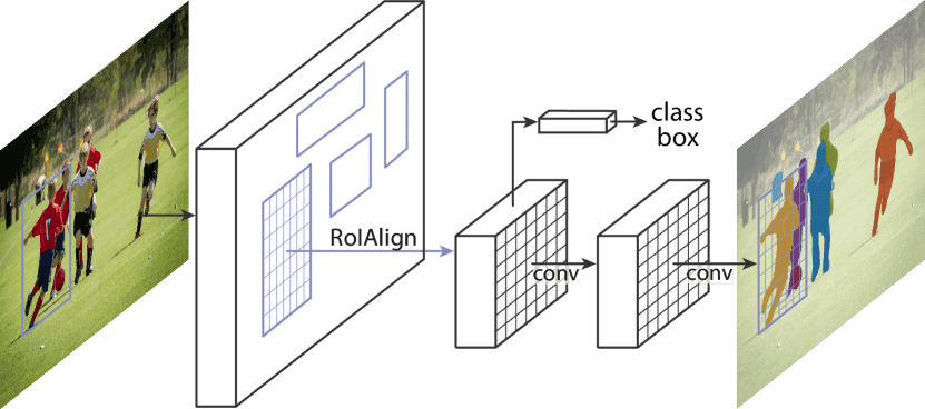 Mask R-CNN: instance segmentation as detection plus one branch
How adding a single mask-prediction branch onto an existing object detector turned 'find the objects' into 'find and precisely outline every object' — and the alignment fix that made small-object masks accurate.
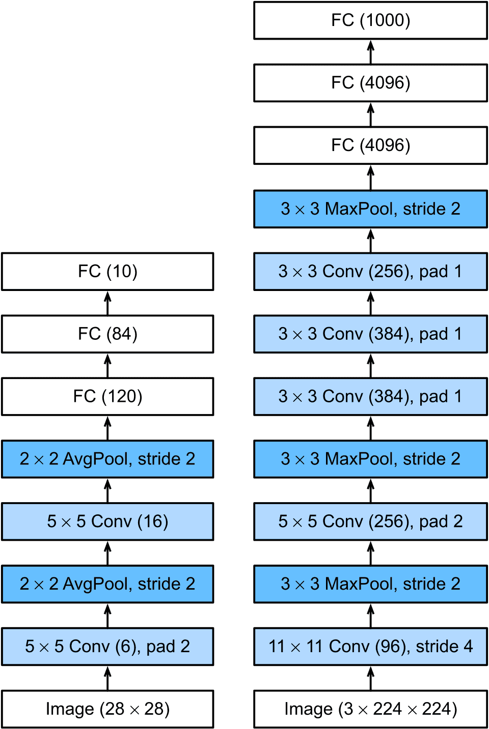 AlexNet: the result that ended the debate about deep learning
How a convolutional network trained on two GPUs cut ImageNet error by a margin large enough to reset computer vision, and why every microscopy segmentation model on this site descends from it.
 Cellpose: segmentation that generalises because it doesn't assume a shape
Why predicting a flow field toward each object's centre, instead of a mask directly, let one trained model segment cell types it had never seen — without retraining.