Digital pathology
Whole-slide H&E image analysis and OpenCV pipelines to detect lymphoid follicles in lymphoma tissue, the subject of my Master's thesis.
01About me
I am interested in how careful software, physical reasoning, and quantitative imaging can make difficult measurements more useful.
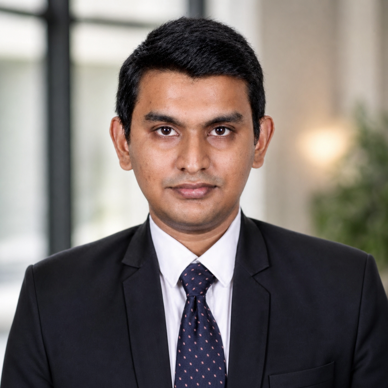
I am currently an Ingénieur Informatique at Phasics (Exosens Group) in Paris, France, a position I began on 15 April 2026. I design and integrate machine learning and computer-vision methods for quantitative phase imaging and scientific instrumentation, building data-processing pipelines that stay robust, scalable, and interpretable in real-world use.
I completed my PhD in Biophysics at Aix-Marseille University on 16 September 2025, jointly affiliated with the Adhesion and Inflammation Laboratory (INSERM U1067 / CNRS UMR 7333) and the Laboratoire d'Informatique et des Systèmes (UMR 7020). My thesis, "Deep Learning-Based Counting and Kinetic Analysis of Bacteriophages from Fluorescence Microscopy," developed a custom TensorFlow architecture for instance-level detection, counting, and localisation of phage-antibody binding events under weak supervision, using TIRF microscopy of antibody-antigen interactions.
The work combined physics-based synthetic datasets grounded in biophysical modelling, domain adaptation across biological conditions and concentration gradients, and physics-informed validation using Poisson statistics, kinetic equations, and simulation models, trained on GPU-accelerated HPC infrastructure with SLURM.
My wider work spans Raman and FTIR spectroscopy, digital pathology, medical imaging, scientific interfaces, and reproducible software. Alongside the research, I have enjoyed contributing to science communication and outreach through scientific cartooning, public talks, and live experiments. I enjoy building tools whose behaviour can be understood, not only benchmarked.
Download complete CV ↓My imaging journey
Whole-slide H&E image analysis and OpenCV pipelines to detect lymphoid follicles in lymphoma tissue, the subject of my Master's thesis.
01Image preprocessing, hotspot analysis, particle localisation, segmentation, and quantitative visualisation for biological data.
02Density-map prediction and single-bacteriophage counting close to the surface, linked to binding kinetics and statistical validation.
03Scientific software and computer-vision work with label-free QPI data, focused on robust and meaningful image-based measurement.
04What I bring
Fluorescence, TIRF, QPI, digital pathology, whole-slide images, and instrument-derived data.
Preprocessing, segmentation, detection, density maps, classification, domain adaptation, and feature engineering.
Statistical modelling, uncertainty-aware evaluation, physical interpretation, controlled data, and reproducibility.
Python, C++, PyTorch, TensorFlow, OpenCV, Qt/QML, HPC/SLURM, Git, and maintainable scientific interfaces.
Timeline
Phasics (Exosens Group) · Saint-Aubin, France
Aix-Marseille University (LAI · LIS) · Thesis defended 16 September 2025
Phoenix Medical Systems · Chennai, India
Grey Scientific Labs · Digital pathology, India
Sri Sathya Sai Institute of Higher Learning · India