Field note
µSAM: closing the gap between SAM and real microscopy images
SAM was trained on natural photographs and performs noticeably worse on fluorescence and electron microscopy out of the box. µSAM fine-tunes it for exactly those modalities and ships it as a usable napari tool.
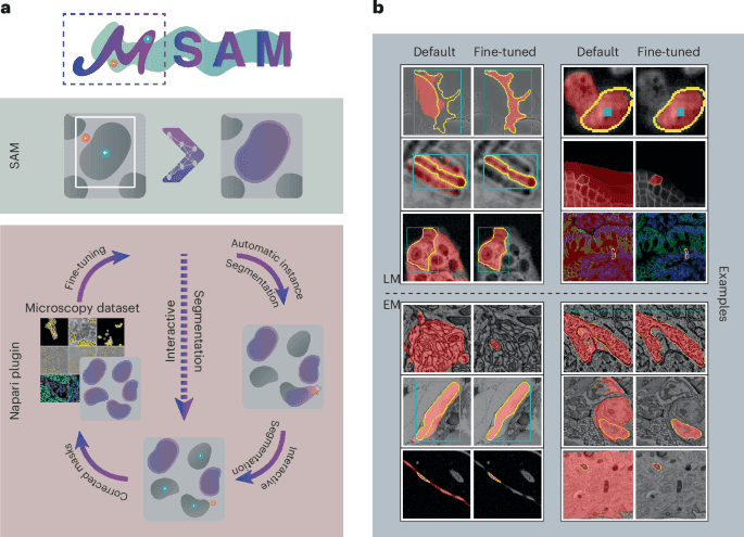
The original SAM’s zero-shot performance on microscopy is real but weaker than on natural photographs — cell boundaries, faint fluorescence signal, and electron-microscopy contrast don’t look like anything in its training data. µSAM (Segment Anything for Microscopy) closes that gap directly: it fine-tunes SAM specifically for light and electron microscopy, then packages the result as annotation, instance-segmentation, and tracking workflows inside napari — the tool working microscopists already use, not a standalone research script.
Fine-tuning, not training from scratch
µSAM starts from SAM’s pretrained weights and adapts them on annotated microscopy datasets, rather than discarding the general-purpose pretraining and starting over. This preserves what SAM already does well — flexible, promptable segmentation from a point or box, no per-object retraining needed — while correcting specifically for the domains it performed worst on. That’s a meaningfully cheaper path than collecting a microscopy-scale SA-1B and training a foundation model from zero.
Shipped as a tool, not just a checkpoint
The distinguishing feature of µSAM isn’t only the fine-tuned weights — it’s the napari integration for interactive annotation, instance segmentation, and object tracking, built around those weights. A fine-tuned model that only exists as a downloadable checkpoint still requires someone to write the annotation UI, the tracking logic, the workflow around it before a working microscopist can use it day to day. µSAM did that integration work as part of the release.
The timeline from submission to publication
Submitted in August 2023, accepted in November 2024, and published in February 2025 — an 18-month review cycle that’s worth noting on its own: the gap between “the underlying method (SAM) was announced” (April 2023) and “a validated, published, microscopy-ready adaptation existed” (February 2025) was the better part of two years, even for a relatively direct fine-tuning application. Foundation-model adaptation to a new scientific domain is not a same-week exercise, however fast the base model’s own announcement was.
Takeaways
- µSAM answers the exact limitation noted in SAM’s own field note: out-of-the-box performance on scientific imaging is weaker than on natural photos, and this is the direct microscopy-specific fix.
- Fine-tuning SAM’s existing weights, rather than training from scratch, is what made this feasible without a microscopy-scale annotated dataset comparable to SA-1B.
- Shipping as an integrated napari workflow, not just model weights, is what makes it a usable tool rather than a research artefact requiring its own UI to be built.
- The 18-month gap between SAM’s release and µSAM’s publication is a realistic benchmark for how long validated domain adaptation of a foundation model actually takes.
Source
Archit et al., Segment Anything for Microscopy, Nature Methods, February 2025 — paper.
Continue through the concepts
Related notes
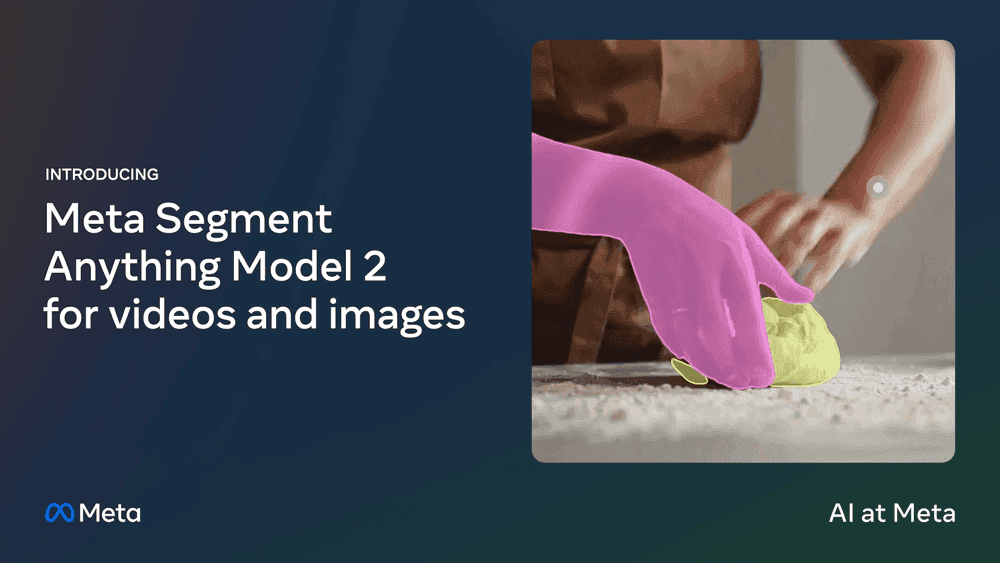 SAM 2: keeping a mask's identity across a video
How a streaming-memory architecture let promptable segmentation extend from single images to video, tracking an object's identity through occlusion and motion in real time.
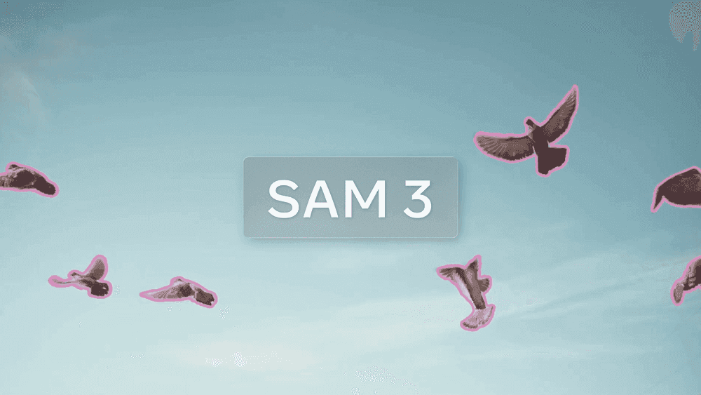 SAM 3: segmenting by concept, not by click
How moving from single-instance prompts to open-vocabulary concept prompts let one phrase or example image segment every matching object in a scene at once.
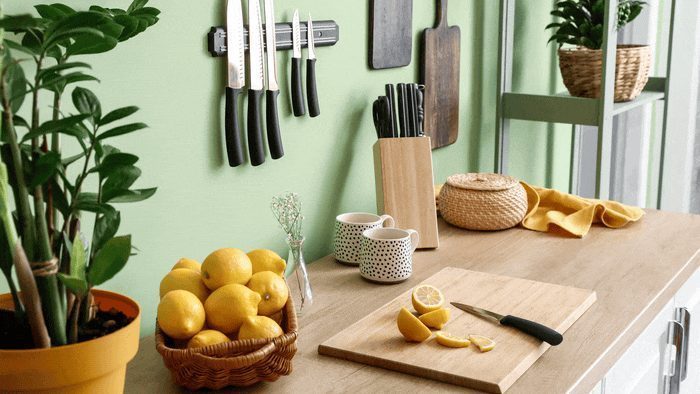 Segment Anything (SAM): segmentation you can prompt instead of retrain
How SAM turned segmentation into a promptable task, why its billion-mask training set exists, and why that idea mattered enough to spawn a whole branch of microscopy-specific models.
 AlexNet: the result that ended the debate about deep learning
How a convolutional network trained on two GPUs cut ImageNet error by a margin large enough to reset computer vision, and why every microscopy segmentation model on this site descends from it.